|
Research Ideas and Outcomes :
Research Idea
|
|
Corresponding author: Yuichi Kano (kano@species.jp)
Academic editor: Daniel Mietchen
Received: 26 May 2022 | Accepted: 02 Aug 2022 | Published: 08 Aug 2022
© 2022 Yuichi Kano
This is an open access article distributed under the terms of the Creative Commons Attribution License (CC BY 4.0), which permits unrestricted use, distribution, and reproduction in any medium, provided the original author and source are credited.
Citation:
Kano Y (2022) Bio-photogrammetry: digitally archiving coloured 3D morphology data of creatures and associated challenges. Research Ideas and Outcomes 8: e86985. https://doi.org/10.3897/rio.8.e86985
|
|
Abstract
Morphological data of life forms are fundamental for documenting and understanding biodiversity. I developed a photogrammetry technique for reconstructing the outer coloured morphology of various creatures and published more than 1000 models online (https://sketchfab.com/ffishAsia-and-floraZia). By suspending it with nylon fishing line(s), taking digital photos from multiple angles and analysing the photos with photogrammetry software, we can obtain a fine 3-dimensional (3D) model of a creature. I believe the challenge could contribute to various fields, such as taxonomy, museology, morphology, anatomy, ecology, education, artificial intelligence, virtual reality, metaverse and, eventually, open/citizen science. Herein, I report the idea and achievement, which I have termed “bio-photogrammetry.”
Keywords
3D model, biological specimen, metaverse, museology, photogrammetry, Sketchfab, virtual reality
Overview and background
The morphological data of creatures are fundamental for documenting and understanding biodiversity. As digital technology develops, digital libraries and archives of biological morphology data/databases have become more important and active (
Photogrammetry is the technology of obtaining information about physical objects by analysing photographic images, radiant imagery and other measuring equipment. In various fields of science, photogrammetry has become popular, especially in the remote sensing and architectonics fields; the 3-dimensional (3D) physical structure of geography, landscape and architecture can be digitally reconstructed through photogrammetry technology (e.g.
Recently I developed a photogrammetry technique for reconstructing various creatures’ outer morphology with colour and published more than 1000 models online at Sketchfab https://sketchfab.com/ffishAsia-and-floraZia (e.g. Figs
Movie of the 3D model of the sailfin poacher, Podothecus sachi. Original data: https://skfb.ly/otRSM or https://ffish.asia/f/84092. The embedded cube with a color chart indicates 10 mm and is scaled according to the real object. The object colour is not standardised (the colour chart is just a rough indication).
Movie of the 3D model of the long arm octopus, Octopus minor. Original data: https://skfb.ly/os7OA or https://ffish.asia/f/83541.
Movie of the 3D model of the hirsute raspberry, Rubus hirsutus. Original data: https://skfb.ly/o9tQ9 or https://floraZia.com/f/2353.
At the moment, I am mainly focusing on aquatic animals, such as fishes (e.g. Fig.
I believe the trial could contribute to various fields, such as taxonomy, museology, morphology, anatomy, ecology, education, artificial intelligence (AI), virtual reality, metaverse and, eventually, open science. Thus, I herein report my photogrammetry challenge and results, obtained by “bio-photogrammetry.”
Methods
The object is suspended by a nylon fishing line(s) and slowly spun and photos are taken from multiple angles (Fig.
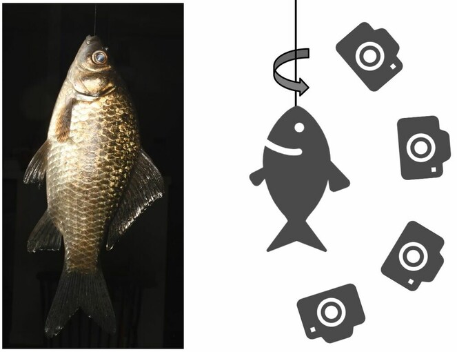
A simplified diagram of the photogrammetry method. The slowly spinning object (Carassius auratus langsdorfii), suspended by a nylon fishing line, is photographed from multiple angles. The resulting 3D model is shown at https://skfb.ly/6ZRDz or https://ffish.asia/f/80064.
There are many photogrammetry software packages available. I use 3DF Zephyr Lite (3Dflow s.r.l., Verona, Italy), but I believe other photogrammetry software would be also suitable for the above protocol provided they accept a high number of photos (at least a few hundred photo images are required to produce an excellent model).
Objectives, significance and application
“Bio-photogrammetry” is especially useful in museology and taxonomy, as the outer morphology of biological specimens can be semi-permanently conserved as digital files. In particular, it is worthwhile 3D modelling valuable or type specimens (e.g. https://skfb.ly/ou8xy). Museums can also contribute to open science by publishing 3D models of their collections (e.g. https://skfb.ly/osGFZ, https://sketchfab.com/search?q=museum&type=users). For taxonomists, the outer coloured 3D model can be published in a paper describing a new species, as well as internal CT/MRI scan data and the DNA sequence.
The models can be also applied to artificial intelligence (AI) that identifies species from a photo image through deep learning (e.g.
3D models are also useful in education (
Issues
The bio-photogrammetry technique has several limitations. For example, transparent creatures, such as some shrimps, cannot be reconstructed. Additionally, reconstruction of eyes, which physiologically absorb light, sometimes fails (e.g. Xylocopa appendiculata circumvolans, Sebastiscus marmoratus). Furthermore, it is almost impossible to make small (< 5 mm) or large (> 1 m) models of objects using this technique (but see
Complex and/or delicate objects are very difficult to reconstruct: thus far, I have failed to produce models of most middle-large sized (< 30–50 cm) herbs with large leaves (but see Arisaema serratum), Comatulida (feather stars) and nudibranchs. However, I believe with further development of software and protocols, these issues will be resolved.
As for CT and/or MRI, "morphosource.org" would be an academically standard repository. However, as far as I know, there is no equivalent repository that archives biological 3D photogrammetry data (while morphosource.org can archive photogrammetry data, the number is still few and they are mostly bones/skulls). Numerous data of biological 3D photogrammetry might exist separately in the respective department or personal websites. As bio-photogrammetry becomes more popular, I expect the emergence of a new online academic repository for the data or the development of the existing platforms (e.g. morphosource.org and Sketchfab), by which the bio-photogrammetry data are made globally standardised, integrated and major.
Impact
In the long history of biology, describing the physical morphology of creatures has advanced gradually from handwritten sketches to digital photo images. Unfortunately, these 2D representations are insufficient for a complete understanding of object morphology. I expect, as a next step, that 3D models produced by “bio-photogrammetry” will enable exciting innovations in various fields.
Data resource
Movies of coloured 3D models of wild creatures made by photogrammetry (Figs
Examples of coloured 3D models of wild organisms made by photogrammetry including the original data of Figs
The author's account of a global platform of the 3D model sharing (
All the data of the author's 3D model of animals (
All the data of the author's 3D model of plants and fungi (
Acknowledgements
This research was partially supported by JSPS KAKENHI JP21H05181 and JP20HP8020.
Ethics and security
This research was conducted ethically under the associated laws. The copyright of all data in this study belongs to the author.
Conflicts of interest
The author declares no conflicts of interest associated with this manuscript.
References
- Library of 3D visual teaching tools for the chemistry classroom accessible via Sketchfab and viewable in augmented reality.Journal of Chemical Education98(9):3032‑3037. https://doi.org/10.1021/acs.jchemed.1c00460
- A new library of 3D models and problems for teaching crystallographic symmetry generated through Blender for use with 3D printers or Sketchfab.Journal of Applied Crystallography55:172‑179. https://doi.org/10.1107/S1600576721013236
- The digital fish library: using MRI to digitize, database, and document the morphological diversity of fish.PloS one7(4):e34499. https://doi.org/10.1371/journal.pone.0034499
- Close-range mini-UAVs photogrammetry for architecture survey.The International Archives of the Photogrammetry, Remote Sensing and Spatial Information SciencesXLII-2:217‑224. https://doi.org/10.5194/isprs-archives-xlii-2-217-2018
- A virtual necropsy: applications of 3D scanning for marine mammal pathology and education.Animals12(4):527. https://doi.org/10.3390/ani12040527
- Unmanned aerial systems for photogrammetry and remote sensing: A review.ISPRS Journal of Photogrammetry and Remote Sensing92:79‑97. https://doi.org/10.1016/j.isprsjprs.2014.02.013
- Database for freshwater fish (+something) biodiversity of Asia. https://ffish.asia. Accessed on: 2022-8-02.
- Database for flora biodiversity of East/SouthEast Asia. https://floraZia.com. Accessed on: 2022-8-02.
- Digital airborne photogrammetry—a new tool for quantitative remote sensing?—a state-of-the-art review on radiometric aspects of digital photogrammetric images.Remote Sensing1(3):577‑605. https://doi.org/10.5167/uzh-24008
- Photo images, 3D models and CT scanned data of loaches (Botiidae, Cobitidae and Nemacheilidae) of Japan.Biodiversity Data Journal6:e26265. https://doi.org/10.3897/BDJ.6.e26265
- Photo images, 3D/CT data and mtDNA of the freshwater mussels (Bivalvia: Unionidae) in the Kyushu and Ryukyu Islands, Japan, with SEM/EDS analysis of the shell.Biodiversity Data ournal7:e32114. https://doi.org/10.3897/BDJ.7.e32114
- Movies of colored 3D models of wild creatures made by photogrammetry. https://doi.org/10.5281/zenodo.6581034. Accessed on: 2022-8-02.
- Colored 3D models of wild creatures made by photogrammetry. https://doi.org/10.5281/zenodo.6577143. Accessed on: 2022-8-02.
- Modeling stratigraphic architecture using small unmanned aerial vehicles and photogrammetry: examples from the Miocene East Coast Basin, New Zealand.Journal of Sedimentary Research87(2):126‑132. https://doi.org/10.2110/jsr.2017.5
- Automatically identifying, counting, and describing wild animals in camera-trap images with deep learning.PNAS115(25):E5716‑E5725. https://doi.org/10.35099/aurora-7
- Application of micro-CT in small animal imaging.Methods50(1):2‑13. https://doi.org/10.1016/j.ymeth.2009.08.007
- Automatic fish species classification in underwater videos: exploiting pre-trained deep neural network models to compensate for limited labelled data.ICES Journal of Marine Science75(1):374‑379. https://doi.org/10.1093/icesjms/fsx109
- ffish.asia / floraZia.com (@ffishAsia-and-floraZia) - Sketchfab. https://sketchfab.com/ffishAsia-and-floraZia. Accessed on: 2022-8-02.