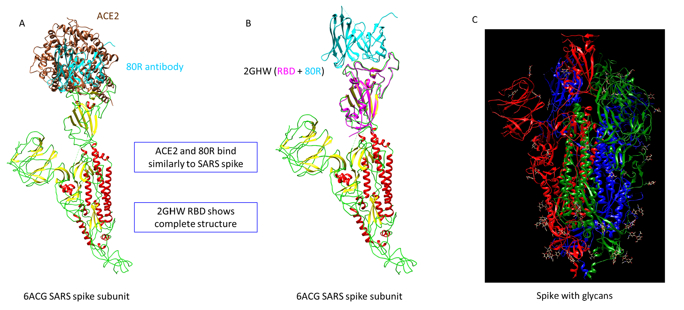
Structural analysis of SARS-CoV spike glycoprotein. In A the SARS-CoV spike protein (PDB ID: 6ACG) is shown bound to ACE2 (brown) and 80R antibody (cyan), superimposed on the same binding site. In B the spike protein is shown bound only to the 80R antibody (PDB ID: 2GHW), with the structural model of the RBD of the SARS-CoV-2 spike protein (magenta) containing the missing loops. This homology model served as the basis for the docking experiments. In C it is shown a spike colored by subunit and showing the glycans. There are only two possible glycans in RBD region at 331 and 343 and neither of these sites affect the 80R binding.