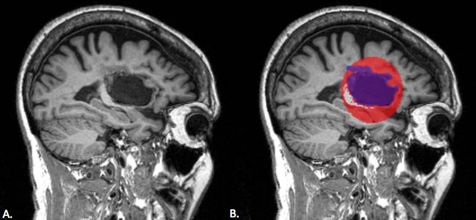
We tested our toolbox on a mock lesion mask. A. The stroke subject's T1 anatomical scan; B. The mock lesion mask is the red sphere; the blue mask is the lesion segmentation. The white matter was intentionally covered within the mock lesion mask, but as shown here, white matter voxels are removed by the white matter correction.